
The most important ophthalmology research updates, delivered directly to you.
The most important ophthalmology research updates, delivered directly to you.
Audio link:
https://open.spotify.com/episode/7o37gwoZDkHNv8MPsibxV6?si=YjCvKH7rRKmObHrd-mzmFw
In this week’s issue
Ophthalmology
Ophthalmic drug shortages in the United States
Why is that drop out of stock?! Medication shortages, both across medicine and in ophthalmology, are frequent and frustrating obstacles for providers and patients alike. Researchers used data from the University of Utah Drug Information Service to identify 379 ophthalmic drugs among 3,089 medications with shortages reported between 2001 and 2024. Systemic and locally delivered medications were represented equally, and most shortages were of anti-infective medications (35%) and steroids (26%). The leading causes of ophthalmic medication shortages were unknown (63%), manufacturing problems (20%), and supply-and-demand mismatches (10%). The median duration of ophthalmic medication shortages was 326 days, nearly 30 days longer than the median of 298 days for all drugs. Mean shortage duration of ophthalmic medications that were patent-protected during each shortage was significantly less (184 days) than medications that experienced a shortage while off-patent (359 days). The majority of ophthalmic medication shortages of “unknown” etiology suggest a troublesome lack of transparency in the drug supply chain and constitute an important target for future quality improvement efforts. The overrepresentation of ophthalmic drugs among the total population of drugs in short supply, as well as the longer mean shortage duration for ophthalmic medications relative to medications overall, highlights the outsized importance of improved drug supply chains for eye health specifically.
JAMA Ophthalmology
Seeing the B3-nefit. Nicotinamide (vitamin B3) has been shown to confer neuroprotective effects in patients with glaucoma. However, it is unknown whether nicotinamide can prevent conversion from ocular hypertension (OHT) to primary open-angle glaucoma (POAG). This retrospective cohort study used the TriNetX federated research network to compare 1460 propensity-score matched patients with OHT exposed to systemic nicotinamide with 1460 matched controls. Over a mean follow-up of 3.7 years, a POAG diagnosis occurred in 3.5% of the nicotinamide group compared with 9% in controls. In addition, nicotinamide exposure was associated with lower rates of initiation of topical IOP-lowering therapy (13.6% vs 21.2%) and laser trabeculoplasty (0.8% vs 1.9%). The protective association of nicotinamide exposure remained consistent in sensitivity analyses of incident users (patients starting nicotinamide only after their OHT diagnosis) and patients with sustained documented use (defined as ≥3 documented prescriptions). These results suggest that systemic nicotinamide may be a complementary strategy for preventing OHT-to-POAG conversion; however, further prospective randomized trials are needed to confirm efficacy and determine the optimal dosing.
American Journal of Ophthalmology
Risk of developing dry eye disease in patients with chronic pain conditions
Is dry eye just the tip of the pain iceberg? Dry eye disease (DED) is increasingly recognized as more than an ocular surface disease, with inflammation and neurosensory dysfunction contributing to symptoms. Growing evidence supports an association between ocular discomfort and chronic pain conditions (CPCs) such as fibromyalgia, migraine, and irritable bowel syndrome, but whether CPCs predict the future development of incident DED remains unclear. This large retrospective propensity score-matched cohort study compared 538,364 adults with at least one CPC to 538,364 controls without any CPC. Patients with CPCs had a 5-fold higher risk of incident DED over 3 years, with cumulative DED rates of 7.16% versus 1.50% in controls. They were more than twice as likely to require escalation to prescription dry eye therapy (cyclosporine or lifitegrast) by 3 years (0.79% versus 0.30%). These findings were consistent in a validation cohort limited to patients with age-related cataracts. Overall, CPCs appear to be a strong independent predictor of future DED, supporting the concept that dry eye may reflect broader abnormalities in pain processing rather than solely localized ocular surface pathology.
British Journal of Ophthalmology
The vitreous humor is no laughing matter when it comes to diabetic retinopathy. Severe non-proliferative diabetic retinopathy (sNPDR) is a pivotal stage of diabetic eye disease with a high risk of progression to high-risk proliferative diabetic retinopathy (PDR), which can lead to vitreous hemorrhage and permanent vision loss. The vitreous humor plays an important role in promoting angiogenesis and inflammation that contribute to neovascularization. Therefore, it has been hypothesized that directly targeting the vitreous with pars plana vitrectomy (PPV) may reduce the risk of progression to PDR and yield better outcomes than the current standard of care, panretinal photocoagulation (PRP). In this randomized controlled trial, 55 eyes from patients with sNPDR were randomized to undergo PPV (n=27) or PRP (n=28) to compare the proportion of eyes that progressed to PDR over 12 months. No eyes in the PPV group progressed to PDR, compared with 2 of 28 eyes in the PRP group, although this difference was not statistically significant. When visual field outcomes were assessed, the PPV group had significantly better preservation of total visual field point score than the PRP group (−16.70 vs. −314.00 dB). As an exploratory outcome, the authors also found a trend toward better diabetic macular edema (DME) outcomes with PPV, with more patients experiencing DME improvement (60% vs. 22%) and fewer experiencing worsening (30% vs. 56%) compared with PRP. Overall, these findings suggest that while PPV did not significantly reduce progression to PDR compared with PRP during the 12-month follow-up period, it was associated with better preservation of peripheral visual function and a favorable trend in DME outcomes.
Eye
Seeing double? Ocular migraines may share a linked risk of stroke
Is your vision playing tricks on you, or is your cardiovascular system trying to send a warning sign? While the link between classic migraines and stroke is well-established, the specific cerebrovascular risks for patients diagnosed with ocular migraine (defined as meeting migraine criteria alongside temporary monocular visual disturbances) have remained largely unexamined. To address this gap, researchers conducted a retrospective cohort study using the TriNetX platform to analyze electronic health record data from 21,949 patients aged 50 and older with an ICD code for ocular migraine, and compared them with matched ophthalmology, migraine with aura (MA), and migraine without aura (MO) controls. Compared to general ophthalmology controls, patients aged 50 and older with ocular migraine had a 48% increased risk of combined cardiovascular and cerebrovascular outcomes (RR 1.48), including a 2.68 times higher risk of transient cerebral ischemic attacks and an 81% increased risk of cerebral infarcts, though their risk profile remained highly comparable to patients with MA and MO. These findings indicate that an ocular migraine diagnostic code carries a vascular risk profile similar to other migraine subtypes, highlighting the need for clinicians to monitor these patients closely and for future prospective studies to confirm the underlying mechanisms.
JAMA Ophthalmology
A smart(phone) approach to ocular malignancy screening
Ocular oncology screening may be getting a pocket-sized upgrade. Basal cell carcinoma and malignant melanoma, among other ocular surface malignancies, pose a significant threat to vision and ocular health. These conditions are frequently dismissed by patients as normal age-related changes and misdiagnosed by non-specialist clinicians, both because of clinical overlap with benign conditions and limited access to specialists trained in ocular oncology. This prospective, three-stage, nonrandomized trial aimed to develop and externally validate CaptureTumor (CaT), a smartphone application for detecting rare ocular surface malignancies. In stage 1, investigators trained a convolutional neural network (CNN) on 12 years of slit-lamp images sourced from four hospitals and multiple public databases. In stage 2, they adapted the model for smartphone photography, integrating it into CaT to facilitate patient use. Stage 3 tested the platform in the real world, reaching over 256,000 individuals. The application analyzes a patient-submitted image and classifies the lesion as either benign or malignant. A total of 614 participants completed at-home screening, and the application demonstrated strong diagnostic performance, achieving an AUC of 0.977, sensitivity of 89.3%, and specificity of 95.9%. Twenty malignancies were confirmed by biopsy, including 14 basal cell carcinomas and 6 malignant melanomas; none of the affected patients required enucleation or orbital exenteration. By expanding access to a smartphone-based, user-friendly screening platform, CaptureTumor may facilitate earlier detection of suspicious ocular lesions and more timely referral for specialist evaluation.
JAMA Network Open
Deep learning predictions of childhood myopia prediction
Childhood myopia might show signs early. Childhood myopia is a rapidly escalating global health concern, with high myopia projected to affect more than 1 billion individuals by 2050 and carrying a substantial risk of myopic maculopathy and permanent vision loss. This longitudinal cohort study from Beijing Tongren Eye Center and Beihang University followed 3,048 grade 1 schoolchildren over 6 years as part of the Anyang Childhood Eye Study, generating 16,211 fundus images to develop and validate a novel deep learning model for myopia progression prediction. The Multiyear Myopia Prediction Network combined a ResNet34 convolutional neural network with a long short-term memory recurrent network, requiring only fundus images and baseline spherical equivalent refraction as inputs. The model achieved an AUC of 0.941 (95% CI, 0.936–0.946) for myopia risk prediction and 0.985 (95% CI, 0.982–0.988) for high myopia risk prediction, with an overall mean absolute error (MAE) of 0.322 D per year for quantitative refraction prediction. For 1-year predictions, AUC reached 0.973 for myopia and 0.999 for high myopia. The model performed better in initially nonmyopic children (MAE 0.44 D/year) than in children with baseline myopia (MAE 0.58 D/year). A deep learning model that predicts high myopia risk with an AUC above 0.98 using nothing more than a fundus photo and a baseline refraction reading has meaningful practical implications, particularly given that pharmacologic myopia control can reduce progression by more than 70% when started early enough.
In the days before anti-VEGF, what did retina docs do? This 1984 trial sought to answer three questions when it comes to managing patients after branch vein occlusion. 1) Can photocoagulation prevent the development of neovascularization (NV)? 2) Can photocoagulation prevent vitreous hemorrhage (VH)? 3) Can photocoagulation improve visual acuity in eyes with macular edema, reducing vision to 20/40 or worse? To do so, 502 eyes were categorized into four groups: I. eyes at risk for development of NV; II. eyes at risk for development of VH; III. Eyes at risk for vision loss; X. eyes at high risk for NV.
Key Points:
Overall, the BVOS is a landmark study because it highlighted the important role of argon laser photocoagulation in the prevention of the development of neovascularization and vitreous hemorrhage in eyes with branch vein occlusion. Further, treatment with argon laser photocoagulation is recommended for those with branch vein occlusion and visual acuity reduced from macular edema to 20/40 or below. With treatment, improvement of visual acuity is more likely than without laser treatment.
When the fistula closes but the vision does not improve
Journal: Case Reports in Ophthalmology
Can corticosteroids improve visual outcomes after successful treatment of a carotid-cavernous fistula? Carotid-cavernous fistulas (CCFs) are vision-threatening vascular abnormalities usually managed with endovascular embolization. Closure of the fistula often resolves symptoms, but some patients may continue to experience persistent orbital inflammation, ophthalmoplegia, and visual decline. The role of corticosteroids in this setting remains poorly defined.
In this case report, a patient with a CCF underwent multiple endovascular embolization procedures to achieve fistula closure. However, despite successful surgical treatment, the patient continued to experience persistent proptosis, ophthalmoplegia, and worsening vision. High-dose intravenous corticosteroids were initiated, resulting in marked improvement in visual acuity, extraocular motility, and orbital congestion. Following the steroid taper, the patient had recurrent chemosis and conjunctival injection that resolved with reinitiation of steroids and a slower taper. The patient's recovery in the setting of steroid initiation suggested that the patient’s symptoms were primarily due to persistent inflammation rather than incomplete CCF closure.
This case highlights the potential role of high-dose corticosteroids as an adjunctive therapy for patients with persistent inflammation following successful CCF closure. Although endovascular embolization remains the mainstay of treatment, recognizing inflammation as a contributor to delayed visual recovery and addressing it with steroids may improve visual outcomes.
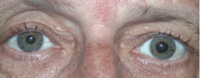
You are the resident on call when a 72-year-old man presents with complaints of glare and a dilated left pupil. One week ago, he underwent uncomplicated cataract surgery on the same eye. Postoperative care included topical antibiotics and anti-inflammatory drops. On postoperative day 1, his left pupil was noted to be 7 mm compared to 3 mm on the right. He reports no pain, and there was no corneal edema or elevated intraocular pressure. Examination now reveals a round, fixed dilated pupil that fails to constrict to topical 2% pilocarpine. Weeks later, iris atrophy is noted.
What is the most likely diagnosis?
A. Horner syndrome
B. Urrets-Zavalia syndrome
C. Pharmacologic mydriasis
D. Intraoperative iris sphincter damage
Follow us on Twitter @TheLens_Oph and Instagram @TheLens_Oph
Contact us if you have inquiries for advertisements, questions, or general feedback for The Lens – we’re always looking to improve!
